

Product Code: 10.7030
Neutron
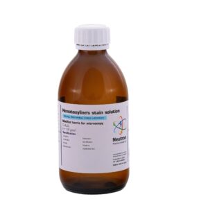
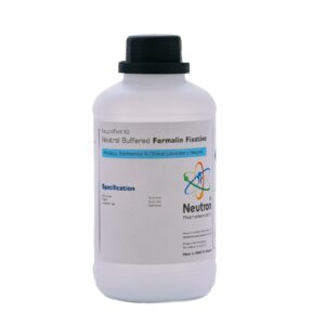
Mallory s stain solution
Formula
Molar Mass
CAS Number
HS Code
Uses
Mallory’s Stain Solution is a histological staining solution widely used for differentiating connective tissues in microscopic examinations. The stain consists of a combination of acidic dyes that selectively color collagen fibers, cytoplasm, muscle fibers, and other tissue components. Mallory’s staining methods are commonly applied in pathology, histology, and biological research laboratories.
🏭⚗️ Synthesis
Mallory’s Stain Solution is prepared by dissolving and combining specific dyes such as aniline blue, acid fuchsin, and orange G in aqueous or acidic solutions under controlled laboratory conditions. Additional reagents, including phosphomolybdic acid, may be incorporated to enhance selective tissue staining and color differentiation.
🧪 Applications
Mallory’s Stain Solution is widely used in histology and pathology for staining connective tissues and differentiating collagen fibers from muscle and cellular structures. It is commonly applied in microscopic examination of biological tissues for diagnostic and research purposes. The solution is also utilized in educational and biomedical laboratories for tissue structure analysis and histological demonstrations.
⚠️ Safety
Mallory’s Stain Solution may cause irritation to the eyes, skin, and respiratory tract due to the presence of acidic dyes and chemical reagents. Prolonged exposure or ingestion should be avoided. Appropriate laboratory protective equipment, including gloves, goggles, and proper ventilation, is recommended during handling and preparation.